Serial millisecond crystallography (SMX) using a syringe pump was performed on the microfocusing beamline 11C at Pohang Light Source II (Park et al., 2017). The X-ray beam size focused by a Kirkpatrick–Baez mirror was 4.1 µm (vertical) × 8.5 µm (horizontal) at the sample position. The and beam energy were 1.3 × 10¹² photons s−1 and 12.659 keV, respectively. The crystallization of lysozyme from chicken egg white was performed using a previously reported procedure (Lee et al., 2019) The lysozyme crystals were embedded in the sample delivery medium (LCP or PAM) using mechanical mixing in a dual-syringe setup.
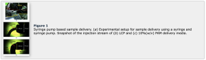
Figure 1
Syringe pump based sample delivery. (a) Experimental setup for sample delivery using a syringe and syringe pump. Snapshot of the injection stream of (b) LCP and (c) 10%(w/v) PAM delivery media.
A 100 µl syringe (Hamilton, 81065-1710RNR) containing the crystals embedded in the delivery medium was mounted on a Fusion Touch 100 syringe pump (CHEMYX). The syringe needle with an inner diameter of 168 µm was monitored by an on-axis video microscope installed in the MD2-S goniometer (Arinax) in the beamline hutch [Fig. 1 (a)]. To align the syringe needle on the Chemyx Fusion 100 syringe pump to the X-ray beam path, a manual stage was installed under the syringe pump stand to make fine adjustments in the x, y and z directions. Due to the interference of the beamline equipment and the syringe pump, the syringes and needles were installed horizontally [Fig. 1(a)]. Considering the stream direction of the delivery medium from the syringe needle, the needle end of the syringe was aligned at approximately 100 µm from the beam path, 45° diagonally upward using manual stages [Figs. 1(b) and 1(c)]. By driving the syringe pump, LCP- and PAM-containing crystals were extruded from the syringe needle to the X-ray position at flow rates of 100 nl min−1 and 200 nl min−1, respectively [Figs. 1(b) and 1(c)].
During data collection, all images were exposed for 100 ms. The temperature and humidity inside the hutch were 25 ± 0.4°C and 20%, respectively. Images were pre-filtered using Cheetah (Barty et al., 2014) and processed using CrystFEL (White et al., 2016). Molecule replacement was performed using Phaser-MR (Adams et al., 2010) with lysozyme (PDB code 6irj; Lee et al., 2019) as the search model. Model building and were carried out using Coot (Emsley & Cowtan, 2004) and Phenix.refinement in PHENIX (Adams et al., 2010), respectively. The geometries were analyzed using MolProbity (Williams et al., 2018). The figures were generated by PyMOL (https://pymol.org/). The coordinates and structure factors have been deposited in the Protein Data Bank under the accession codes 6JXP (LCP-lysozyme) and 6JXQ (PAM-lysozyme). Diffraction images have been deposited in CXIDB under IDs 95 (LCP-lysozyme) and 96 (PAM-lysozyme).
Authors: Suk-Youl Parka and Ki Hyun Namb
Date Published: September 2019
Read full article here: Sample delivery using viscous media, a syringe and a syringe pump for serial crystallography